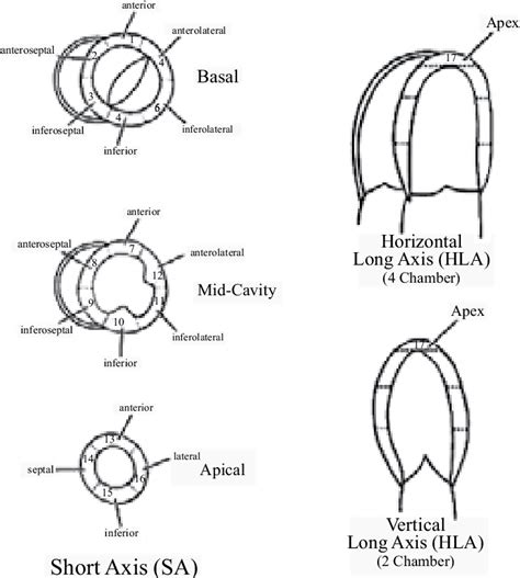
lv hypokinesis Mild hypokinesia basically means that the muscle of your heart does not contract as much as most peoples' hearts do. This may sound scary, but, do not be too worried because your ejection. There's an issue and the page could not be loaded. Reload page. 2,470 Followers, 188 Following, 539 Posts - See Instagram photos and videos from Empower LV (@empower.lv)
0 · what is mild septal hypokinesis
1 · what does severely hypokinetic mean
2 · severe hypokinesis of the apex
3 · severe hypokinesis of left ventricle
4 · mild apical and septal hypokinesis
5 · hypokinesis of left ventricle cause
6 · hypokinesis of heart wall treatment
7 · causes of septal hypokinesis
Epic Real Estate, led by Matt Theriault, is a community and set of tools and resources for real estate investors. If you want to build a steady stream of passive income by investing in real estate, then Epic Real Estate is designed specifically for you.
Mild hypokinesia basically means that the muscle of your heart does not contract as much as most peoples' hearts do. This may sound scary, but, do not be too worried because your ejection.

Global hypokinesis of the heart is a condition characterized by the weakening of the heart muscle, leading to reduced heart function. This condition can result in serious health .Left-sided heart failure occurs when the heart loses its ability to pump blood. This prevents organs from receiving enough oxygen. The condition can lead to complications that include .
Echocardiographic evaluation of wall motion (WM) is a simple, well-validated method to assess segmental left ventricular (LV) function. 1,2 .
The prognosis of left ventricular noncompaction (LVNC) remains elusive despite its recognition as a clinical entity for >30 years. We sought to identify clinical and imaging characteristics and risk factors for mortality in . The most common wall motion abnormality in NSC due to SAH appears to be either hypokinesis of the basal and midventricular segments with sparing of the apex or global LV hypokinesis, whereas the TC pattern is less .Herein we review the conventional assessment of LV systolic function and examine the role of speckle-tracking echocardiography (STE), a new method to assess LV function. We also highlight the role of STE in the assessment and .
Segmental LV motion abnormalities in the form of hypokinesis in patients with CAD are associated with the presence of embolic signals in the middle cerebral arteries, which may . We can see some hypokinesis of the anterior wall and overall mildly reduced heart function and ejection fraction. A reduced heart function and ejection fraction (EF) (<40%) usually manifests as fatigue and shortness of . The left ventricle (LV) does not empty out with each contraction. Normally the left ventricle (LV) ejects between 50% and 70% of the blood it contains. Below is an echocardiogram of a patient with a normal ejection .
Despite the many advances in cardiovascular medicine, decisions concerning the diagnosis, prevention, and treatment of left ventricular (LV) thrombus often remain challenging. There are only limited organizational . Furthermore, it is unclear whether LV wall motion abnormality is independently associated with embolic stroke. 5 Whereas LV akinesia or hypokinesis were previously considered risk factors for cardioembolism, . Cardiac wall motion abnormalities describe kinetic alterations in the cardiac wall motion during the cardiac cycle and have an effect on cardiac function.Cardiac wall motion abnormalities can be categorized with respect to their degree and their distribution pattern that is whether they are global or segmental and whether they can be attributed to a coronary .Your left ventricle is the largest and strongest chamber of your heart.It’s responsible for pumping oxygen-rich blood from your lungs to the rest of your body. When the left ventricle is weak it can cause fluid to build up in your lungs, resulting in shortness of breath or fatigue.
LV diastolic dysfunction is a feature of HF, both with preserved and with reduced ejection fraction. We were unable to evaluate the separate and distinct contribution of diastolic dysfunction to HF as serial diastolic function parameters were unavailable at the baseline examination we chose. However, it is often challenging to differentiate the . Absolutely in all patients who suffered a myocardial infarction, subsequently on a cardiogram they detect hypokinesia of the heart. As a rule, this happens about two months after the infarction.
what is mild septal hypokinesis
There are many disorders that may involve the left ventricular (LV) apex; however, they are sometimes difficult to differentiate. In this setting cardiac imaging methods can provide the clue to obtaining the diagnosis. The purpose of this review is to illustrate the spectrum of diseases that most frequently affect the apex of the LV including Tako-Tsubo cardiomyopathy, . Hypokinesis was classified as global when it symmetrically involved all segments or segmental if it was predominantly localized to specific segments. . LV ejection fraction was also entered into the regression model that accounted for gender, age, waist-hip ratio, systolic blood pressure, diabetes mellitus, and segmental WM abnormalities. . Hypokinesis is a condition characterized by reduced movement or contraction of the heart muscle, impacting its ability to pump blood effectively. This phenomenon often manifests in various cardiac disorders, including myocardial infarction, cardiomyopathy, and heart failure. Understanding this condition is crucial as it can significantly impact . It may also identify the most common/typical pattern of Takotsubo, which has apical and mid-ventricular wall hypokinesis with basal hyperkinesis. 29 Atypical variants include circumferential mid-ventricular wall hypokinesis with basal and apical hyperkinesis, reverse Takotsubo with isolated basal hypokinesis, focal segmental LV hypokinesis and .
designer inspired handbags wholesale
The LVEF refers to the percentage of blood that is pumped out of the left ventricle with each heartbeat. The LVEF for a healthy heart is between 55% and 70%. The LVEF may be lower if your heart has been damaged. Echocardiography is .We would like to show you a description here but the site won’t allow us.
Uncontrolled high blood pressure is the most common cause of left ventricular hypertrophy. Complications include irregular heart rhythms, called arrhythmias, and heart failure.
harga parfum burberry
what does severely hypokinetic mean
Bouthoorn S, et al. (2018). The prevalence of left ventricular diastolic dysfunction and heart failure with preserved ejection fraction in men and women with type 2 diabetes: A systematic review . Johnson Francis. Former Professor of Cardiology, Calicut Govt. Medical Kozhikode, Kerala, India. Editor-in-Chief, BMH Medical JournalAn echocardiogram revealed moderate-to-severe global hypokinesis of the LV, ejection fraction (EF) estimated at 30%, a 19×7 mm thrombus in the LV apex and a mildly dilated left atrium with tissue Doppler features of diastolic dysfunction (figures 1 and 2, video 1). Acute decompensated heart failure (ADHF) responded appropriately to aggressive .

We would like to show you a description here but the site won’t allow us.
In one patient, cardiogenic shock developed only after rupture of the interventricular septum, four subjects had "primary" cardiogenic shock. In these four persons there were found extensive disturbances of left ventricular wall motion (the mean extent of the akinetic or dyskinetic zone amounted to 41% of the left ventricle (LV).
severe hypokinesis of the apex

Level 40: Reach level 40. Well, if you don’t have anything better to do, you might as well go even further. Limits Were Broken: Use an awesome limit break for the first time. Lo And Behold: Unknown. Lost Ruins: Find the hidden ruins in the jungle. Lumberjack: Unknown. Lunchtime: Take a break and have something to eat outside of battle.
lv hypokinesis|causes of septal hypokinesis


