hypokinesia of lv Cardiac MRI showed a moderately dilated LV with mild-to-moderate concentric LV hypertrophy with wall thickness of 13 mm. LV systolic function was severely reduced with quantitative EF of . Bankas konts* IBAN. Konta īpašnieks* BIC numurs* Reģistrācijas apstiprināšanas gadījumā elektroniskās deklarēšanās sistēmas lietotāja vārds un sākotnējā parole tiks nosūtīta uz iesniegumā norādīto epastu. Saglabāt .
0 · what is mild septal hypokinesis
1 · what does severely hypokinetic mean
2 · severe hypokinesis of the apex
3 · severe hypokinesis of left ventricle
4 · mild apical and septal hypokinesis
5 · hypokinesis of left ventricle cause
6 · hypokinesis of heart wall treatment
7 · causes of septal hypokinesis
Gerardo Díaz y su Gerarquia
Cardiac MRI showed a moderately dilated LV with mild-to-moderate concentric LV hypertrophy with wall thickness of 13 mm. LV systolic function was severely reduced with quantitative EF of .Presence of LV akinesia/hypokinesia and/or LVEF < 55 % significantly increased the odds of having ACA in patients undergoing DCC. Currently, LVWM and LVEF are important indicators . Left ventricular dysfunction is the medical name for a weak heart pump. It's a condition that impacts about 9% of people over the age of 60, which is around 7 million .
gucci moccasins
The prognosis of left ventricular noncompaction (LVNC) remains elusive despite its recognition as a clinical entity for >30 years. We sought to identify clinical and imaging characteristics and risk factors for mortality in . Global hypokinesis of the heart is a condition characterized by the weakening of the heart muscle, leading to reduced heart function. This condition can result in serious health .
Diagnoses most commonly associated with LV dysfunction and non-cardiac disease were sepsis, respiratory insufficiency, major haemorrhage, and neurological disorders. RWMA (n = 40) with or without low EF was more .Global left ventricular hypokinesia is very frequent in adult septic shock and could be unmasked, in some patients, by norepinephrine treatment. Left ventricular hypokinesia is usually corrected . Presence of LV wall motion abnormality was similarly based on transthoracic or ‐esophageal echocardiography results, as interpreted by an attending cardiologist, and were defined as hypokinesia or akinesia of 1 or .
Acute left ventricular (LV) dysfunction occurs in about one-third of critically ill hospitalized patients. 1, 2, 3, 4 The increasing incidence of LV dysfunction in ICUs is likely . Left ventricular dysfunction is the medical name for a weak heart pump. It's a condition that impacts about 9% of people over the age of 60, which is around 7 million Americans. In this Mayo Clinic Minute, Dr. Paul Friedman, a Mayo Clinic cardiologist, explains what the condition is and how it can be diagnosed [.]
Ejection fraction (EF) is a percent measurement of how much blood the left ventricle (LV) pumps with each contraction. The left ventricle (LV) does not empty out with each contraction. Normally the left ventricle (LV) .Introduction: Global left ventricular (LV) hypokinesis is considered to be the cause of stroke, while the significance of segmental wall motion abnormalities is still unknown. Objectives: The aim of the study was to determine the frequency of embolic signals in the middle cerebral artery in patients with segmental LV wall hypokinesis in the course of coronary artery disease (CAD) . Cardiac wall motion abnormalities describe kinetic alterations in the cardiac wall motion during the cardiac cycle and have an effect on cardiac function.Cardiac wall motion abnormalities can be categorized with respect to their degree and their distribution pattern that is whether they are global or segmental and whether they can be attributed to a coronary .Hypokinesia. Hypokinesia refers to decreased bodily movement. Hypokinesiat is characterized by slow movement (bradykinesia) or no movement (akinesia) 1. In Parkinson’s disease, hypokinesia co-occurs with tremor at rest and with rigidity.
Furthermore, it is unclear whether LV wall motion abnormality is independently associated with embolic stroke. 5 Whereas LV akinesia or hypokinesis were previously considered risk factors for cardioembolism, recent evidence suggests that LV wall motion abnormality may not be associated with embolic stroke. 5, 6, 7 In order to evaluate whether . Despite transformative ways in which individuals presenting with an acute coronary syndrome (ACS; including acute non‐ST‐segment–elevated and ST‐segment–elevated myocardial infarction [MI]) are now managed with an early revascularization approach to preserve their myocardium and reduce the risk of mortality, coronary artery disease remains a leading .
Echocardiographic evaluation of wall motion (WM) is a simple, well-validated method to assess segmental left ventricular (LV) function. 1,2 The presence of qualitative WM abnormalities has been demonstrated to be an independent predictor of cardiovascular events in groups of patients with myocardial infarction (MI), 3,4 unstable angina, 5 typical chest pain, 6 .

Global hypokinesia of LV क्या होता है l DCMP echo #echo #shorts the upper two chambers are called atria and the lower two are known as ventriclesMuscular wal. What should I do for severe hypokinesia of the inferior and basal septal walls in my cardio exam? What is severe global hypokinesia? What is the management of LV RWMA (inferior wall hypokinesia) with normal ejaculation fraction?? Hypokinesia of Anterior and Lateral walls, does this situation demand operation or is it curable by Medicines?Mild (grade II) LV systolic dysfunction with global hypokinesis is often consistent with a normal myocardium in atrial fibrillation, when the observation has no other meaning unless specific segmental wall motion defects are also identified. In atrial fibrillation, marked bradycardia or tachycardia (data commonly used to assess diastolic .
The heart is comprised of the pericardium, myocardium, and endocardium. Pathology in any of those structures can lead to heart failure. Left ventricular failure occurs when there is dysfunction of the left ventricle causing insufficient delivery of blood to vital body organs. Left ventricular failure can further subdivide into heart failure with preserved ejection . Despite the many advances in cardiovascular medicine, decisions concerning the diagnosis, prevention, and treatment of left ventricular (LV) thrombus often remain challenging. There are only limited organizational guideline recommendations with regard to LV thrombus. Furthermore, management issues in current practice are increasingly complex, including . About Press Copyright Contact us Creators Advertise Developers Terms Privacy Policy & Safety How YouTube works Test new features NFL Sunday Ticket Press Copyright .
What is LV apical septal hypokinesis? By chatting and providing personal info, you . What is LV apicoseptal hypokinesia? What does this mean : The report states good LV and RV function. LV shows apicoseptal hypokinesiawith other wall showing good contractility. Other cardiac chambers and great vessels normal. Left ventricular asynergy, a frequent cause of myocardial dysfunction in subjects with ischemic heart disease, was studied on ciné-ventriculograms obtained before and after direct coronary artery surgery in 41 patients. Following successful aorto-coronary bypass, hypokinesis of the left ventricle is completely reversible in most instances. Akinesis, on the other hand, is not . Tests normal except MRI had LV EF of 43% & mild hypokinesis. Echo 1yr ago was 59%. Cause of drop & serious? Total wall motion score is 1.18. hypokinesis of the basal to mid anteroseptal wall. hypokinesis of the mid inferoseptal wall. The remaining left ventricular segments demonstrate normal wall motion. EF 47%.
Hypokinesia is caused by a loss of dopamine in the brain. Dopamine — a neurotransmitter, which helps your nerve cells communicate — plays an important role in your motor function.
Normal left ventricular size and global systolic function with inferolateral hypokinesia, mitral annulus dilatation, and bileaflet tethering. . even with normal LV geometry. 3 The analysis of mitral leaflet coaptation pattern with leaflet tethering or flattening may provide more clues about MR mechanism and its clinical impact than . The literature on pheochromocytoma-mediated cardiomyopathy, which is largely limited to case reports, indicates that associated cardiac dysfunction is uncommon in the absence of catecholamine crisis and that it resolves after resection of the tumor. 85–90 However, subclinical LV dysfunction is found in those with normal LV ejection fraction .
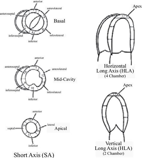
Normal ventricular contraction consists of simultaneous myocardial thickening and endocardial excursion toward the center of the ventricle Regional contractility or wall motion of the LV is graded by dividing the LV into segments In 2002, a 17-segment model was recommended by the American Society of Echocardiography LV is divided into three . Global Hypokinesia of LV l Severe TR l Severe PAH echocardigram #echo test #echocardiography #echocardiogram #ecg By Babita @echocardiographytestnicl5039 .-----A normal LV EF is about 59% to 75%, so the LV EF is a bit less than the usual range Moderate hypokinesis of basal to mid inferior wall ----This indicates the heart muscle at the basal and lower regions of the heart is not moving as much as normally expected, and this may indicate heart attack causing weakness of the heart muscle in .My 26 year old daughter who has Friedreichs Ataxia is in hospital with congestive heart failure she has this report, LV. 12.18.2016. khagihara. Trained in the multiple medical fields for many years. 5,948 Satisfied Customers. Explain what T2/FLAIR hyper-intensities are. Here is the whole report I am reading: PROCEDURE: MRI BRAIN WO CONTRAST
Takotsubo cardiomyopathy (TTC), a syndrome of acute left ventricular (LV) dysfunction, is characterized by transitory hypokinesis of LV apices with compensatory hyperkinesis of the LV basal region. The symptoms of TTC mimic acute myocardial infarction, without significant coronary stenoses on coronary angiography.Ironically, early in the day before i went to hospital I got an Echo cardiogram as i was generally a bit worried about my heart. I got the results on Thursday and it was told my LV ejection fraction was only operating at 30% due to Global Hypokinesis. I have been since taken off Vyvanse and prescribed Entresto and Bisoprolol. Left ventricular dysfunction was defined as having regional wall motion abnormalities (RWMA) or global hypokinesia. RWMA, in turn, was defined as having at least two hypokinetic or akinetic segments with or without an ejection fraction (EF) < 50%. Global hypokinesia was defined as hypokinesia affecting all segments of the LV and an EF < 50%.
what is mild septal hypokinesis
Simplify and reduce the cost of sink installation with Rakks ADA compliant aluminum vanity brackets. These sturdy brackets provide a stable mounting surface for custom-built enclosures and are supplied with wooden strips on the front faces for easy mounti
hypokinesia of lv|what does severely hypokinetic mean


